Кубанов А.А., Абрамова Т.В., Мураховская Е.К., Ласачко В.А. Современный взгляд на лепру. Лечащий Врач. 2018; 5: 48.
Bhandari J., Awais M., Robbins B.A., Gupta V. Leprosy. 2023 Sep 15. In: StatPearls [Internet]. Treasure Island (FL): StatPearls Publishing; 2025 Jan–.
Сароянц Л.В., Арнаудова К.Ш., Дуйко В.В. Идентификация микобактерий, выделенных из содержимого трофических язв больных лепрой. Клиническая дерматология и венерология. 2015; 14 (5): 28–31 .
Белопасов В.В. Типология и патогенез нейропатической боли при лепре. РМЖ. Медицинское обозрение. 2018; (9): 41–45.
Семенова В.Г., Карамова А.Э., Нефедова М.А. и др. Случай погранично-туберкулоидной лепры с атипичными и осложненными проявлениями. Вестник дерматологии и венерологии. 2019; 95 (4): 67–78 .
Henneberg M., Holloway-Kew K., Lucas T. Human major infections: Tuberculosis, treponematoses, leprosy – A paleopathological perspective of their evolution. PLoS One. 2021 Feb 25; 16 (2): e0243687.
Дуйко В.В., Наумов В.З., Сароянц Л.В., Астафурова А.П. Заболеваемость лепрой в прикаспийском регионе. Актуальные вопросы современной медицины. Материалы III Международной конференции Прикаспийских государств. 2018. – С.64–66.
Sharma R., Singh P., McCoy R.C. et al. Isolation of Mycobacterium lepromatosis and development of molecular diagnostic assays to distinguish Mycobacterium leprae and M. lepromatosis. Clin Infect Dis. 2020; 71 (8): e262–e269.
Sotiriou M.C., Stryjewska B.M., Hill C. Two cases of leprosy in siblings caused by Mycobacterium lepromatosis and review of the literature. Am J Trop Med Hyg. 2016; 95 (3): 522–527.
Singh P., Benjak A., Schuenemann V.J. et al. Insight into the evolution and origin of leprosy bacilli from the genome sequence of Mycobacterium lepromatosis. Proc Natl Acad Sci U S A. 2015; 112 (14): 4459–4464.
Образцова О.А. Молекулярно-биологические методы исследования в лабораторной диагностике лепры: эпидемиологический анализ, генетические детерминанты резистентности к антимикробным препаратам. Вестник дерматологии и венерологии. 2017; (6): 34–40.
Kaur G., Kaur J. Multifaceted role of lipids in Mycobacterium leprae. Future Microbiol. 2017; 12 (4): 315–335.
Чеботарев В.В. Тропические болезни в практике дерматовенеролога: учебно-методическое пособие для внеаудиторной работы студентов лечебного и педиатрического факультетов / сост.: В.В. Чеботарев, А.В., Одинец, М.С. Асхаков, Е.В. Бронникова. Ставрополь: Изд-во СтГМУ, 2016, 56 с.
Ploemacher T., Faber W.R., Menke H. et al. Reservoirs and transmission routes of leprosy: A systematic review. PLoS Negl Trop Dis. 2020 Apr 27; 14 (4): e0008276.
Белопасов В.В. Лепра. В кн.: Хронические нейроинфекции / [Баранова Н.С., Белопасов В.В., Бойко А.Н. и др.]; под ред. И.А. Завалишина [и др.]. – 2-е изд. – Москва : ГЭОТАР-Медиа, 2017. – 589 с.
Семенова В.Г., Макарова Л.Л., Карамова А.Э. Узловатая эритема как лепрозная реакция. Вестник дерматологии и венерологии. 2020; 96 (3): 68–74.
Bhat R.M., Prakash C. Leprosy: an overview of pathophysiology. Interdiscip Perspect Infect Dis. 2012;2012:181089.
Scollard D.M., Truman R.W., Ebenezer G.J. Mechanisms of nerve injury in leprosy. Clin Dermatol. 2015; 33 (1): 46–54.
Исмурзаева Н.С. Анализ новых случаев лепры в Астраханской области // Материалы всероссийского научного форума студентов с международным участием «Студенческая наука - 2019». Forcipe. 2019; 2 (S1): 397.
Walker E.L. Some new aspects of the etiology and endemiology of leprosy. Journal of Preventive Medicine. 1929; 3 (3) 167–195.
Mira M.T., Francio, A.S., Sindeaux, R.H. et al. Aspectos geneticos da suscetibilidade do hospedeiro a hanseniase [revisao]. Salud (i) ciencia (Impresa). 2011: 18 (2): 138–141.
Lastória J.C., Abreu M.A. Leprosy: review of the epidemiological, clinical, and etiopathogenic aspects - part 1. An Bras Dermatol. 2014; 89 (2): 205–218.
Pedley J.C. The presence of M. leprae in human milk. Lepr Rev. 1967; 38 (4): 239–242.
Huang C.L. The transmission of leprosy in man. Int J Lepr Other Mycobact Dis. 1980; 48 (3): 309–318.
Колесников Ф.Н. Лепра: из тени на свет. Очерк о загадочной болезни // Universum: Медицина и фармакология: электрон. научн. журн. 2018. № 1 (46). URL: http://7universum.com/ru/med/ archive/item/5443
Янчевская Е.Ю., Меснянкина О.А. Лепра: современные представления о путях передачи. Кубанский научный медицинский вестник. 2019; 26 (4): 123–130.
Geater J.G. The fly as potential vector in the transmission of leprosy. Lepr Rev. 1975; 46 (4): 279–286.
Meredith A., Pozo J.D., Smith S. et al. Leprosy in red squirrels in Scotland. Vet Rec. 2014; 175 (11): 285–286.
Schilling A.K., van Hooij A., Corstjens P. et al. Detection of humoral immunity to mycobacteria causing leprosy in Eurasian red squirrels (Sciurus vulgaris) using a quantitative rapid test. Eur J Wildl Res. 2019; 65: 49.
Kirchheimer W.F. The role of arthropods in the transmission of leprosy. Int. J. Lepr. 1976; 44 (1–2): 104–107.
Tio-Coma M., Wijnands W., Pierneef, L. et al. Detection of Mycobacterium leprae DNA in soil: multiple needles in the haystack. Sci Rep. 2019 Feb 28; 9 (1): 3165.
Mohanty P.S., Naaz F., Katara D. et al., Viability of Mycobacterium leprae in the environment and its role in leprosy. Indian J Dermatol Venereol Leprol. 2016; 82 (1): 23–27.
World Health Organization: Weekly epidemiological record, 30 AUGUST 94th YEAR: Nos.35/36,2019,94,389-412 [Электронный ресурс]. Режим доступа: http://www.who.int/wer (дата обращения: 20.09.21).
Guidelines for the diagnosis, treatment and prevention of leprosy. - ISBN: 978-92-9022-6383/ - World Health Organization. - 2018. - 87 p.
Скрипкин Ю.К., Кубанова А.А., Акимов В.Г. Кожные и венерические болезни. - ГЭОТАР-Медиа, 2012. – 544 с.
Ramos J.M., Romero D., Belinchón I. Epidemiology of leprosy in Spain: the role of the international migration. PLoS Negl Trop Dis. 2016 Mar 3; 10 (3): e0004321.
Aftab H., Nielsen S.D., Bygbjerg I.C. Leprosy in Denmark 1980–2010: a review of 15 cases. BMC Res Notes. 2016 Jan 5; 9: 10.
Massone C., Brunasso A.M., Noto S. et al. Imported leprosy in Italy. J Eur Acad Dermatol Venereol. 2012; 26 (8): 999–1006.
Лепра. Доклад ВОЗ 22 янв. 2010 г. [Электронный ресурс]. Режим доступа: http://apps.who.int/gb/ebwha/pdf_files/EB126/B126_41-ru.pdf (дата обращения: 05 августа 2025 г.).
Ridley D.S., Jopling W.H. Classification of leprosy according to immunity. A five-group system. Int J Lepr Other Mycobact Dis. 1966; 34 (3): 255–273.
Chaitanya V. S., Cuello L., Das M. et al. Analysis of a novel multiplex polymerase chain reaction assay as a sensitive tool for the diagnosis of indeterminate and tuberculoid forms of leprosy. Int J Mycobacteriol. 2017; 6 (1): 1–8.
ВОЗ. Руководство по диагностике, лечению и профилактике лепры. – Женева: ВОЗ, 2018. – 106 с.
World Health Organization Leprosy elimination [cited Apr 2021]; Available from:http://www.who.int/lep/epidemiology/en/.
World Health Organization et al. Лепра: Ведение больных с лепрозными реакциями и профилактика инвалидности. Техническое руководство.
Адаскевич В.П., Козин В.Н. Кожные и венерические болезни. – М.: Медицинская литература, 2006. – 659 с.
Белопасов В.В., Андросюк Ю.Г., Дячина М.Н. Лепрозные невропатии. Журнал неврологии и психиатрии им. C.C. Корсакова. 2004; 104 (11): 19–24.
Андросюк Ю.Г., Дячина М.Н., Белопасов В.В., Корнеева М.А. Клиникодиагностическая оценка лепрозных невропатий в стадии обострения. Нейроиммунология. 2004; (2): 6–7.
Семенова В.Г., Макарова Л.Л., Карамова А.Э. Узловатая эритема как лепрозная реакция. Вестник дерматологии и венерологии. 2020; 96 (3): 68–74.
Upputuri B, Pallapati MS, Tarwater P, Srikantam A. Thalidomide in the treatment of erythema nodosumleprosum (ENL) in an outpatient setting: A five-year retrospective analysis from a leprosy referral centre in India. PLoS Negl Trop Dis. 2020 Oct 9; 14 (10): e0008678.
Talhari C., Talhari S., Penna G.O. Clinical aspects of leprosy. Clin Dermatol. 2015; 33 (1): 26–37.
Lastória J.C., Abreu M.A. Leprosy: review of the epidemiological, clinical, and etiopathogenic aspects - part 1. An Bras Dermatol. 2014; 89 (2): 205–218.
Лезвинская Е.М., Романенко Г.Ф., Казанцева И.А. и др. Случай пограничного типа лепры. Российский журнал кожных и венерических болезней. 2004; (2): 20–22.
Янчевская Е.Ю., Дуйко В.В., Меснянкина О.А. Классификация лепры: исторические аспекты, современный подход. Лечебное дело. 2020; (1): 6–11.
Янчевская Е.Ю., Ковтунова В.А., Думченко Е.В., Дуйко В.В. Погранично- лепроматозная лепра с первичной клинической манифестацией у пациента с длительным дерматологическим анамнезом. Вестник дерматологии и венерологии. 2020; 96 (5): 53–58.
Торсуев Н.А. Распознавание и дифференциальная диагностика лепры. – М.: Медицина, 1971. – 184 с.
Торсуев Н.А. К вопросу о лечении лепры. Вестник венерологии и дерматологии. 1955; 37: 35.
Silva I.M., Oliveira C.A., Guedes W.R. et al. Agranulocytosis induced by multidrug therapy in leprosy treatment: a case report. Braz J Infect Dis. 2009; 13 (2): 158–160.
Guragain S., Upadhayay N., Bhattarai B.M. Adverse reactions in leprosy patients who underwent dapsone multidrug therapy: a retrospective study. Clin Pharmacol. 2017 Jun 29; 9: 73–78.
Deps P.D., Nasser S., Guerra P. et al. Adverse effects from multi-drug therapy in leprosy: a Brazilian study. Lepr Rev. 2007; 78 (3): 216–222.
Satarasinghe R.L., Jayawardana M.A., De Silva G.V. et al. Total agranulocytosis caused by dapsone therapy for tuberculoid leprosy – an unappreciated serious side effect of anti-leprosy treatment with clinical implications. Drug Metabol Drug Interact. 2009; 24 (2–4): 325–329.
Moura A.K., Melo B.L., Brito A.E. et al. High levels of liver enzymes in five patients with type 1 leprosy reaction. J Eur Acad Dermatol Venereol. 2009; 23 (1): 96–98.
Polito M.G., Moreira S.R., Nishida S.K, Mastroianni Kirsztajn G. It is time to review concepts on renal involvement in leprosy: pre- and post-treatment evaluation of 189 patients. Ren Fail. 2015; 37 (7): 1171–1174.
Ozturk S, Ozturk T, Can I. Renal involvement in leprosy: evaluation of patients in Turkey. Postepy Dermatol Alergol. 2017; 34 (3): 240–244.
Логинов В.К. Лепрозное поражение печени: дис. – М.: автореф. дис. д-ра мед. наук, 1969.
Penna G.O., Pontes M.A., Cruz R. et al. A clinical trial for uniform multidrug therapy for leprosy patients in Brazil: rationale and design. Mem Inst Oswaldo Cruz. 2012 Dec;107 Suppl 1: 22–27.
Weiand D., Thoulass J., Smith W.C. Assessing and improving adherence with multidrug therapy. Lepr Rev. 2012; 83 (3): 282–291.
Silva E.A., Iyer A., Ura S. et al. Utility of measuring serum levels of anti-PGL-I antibody, neopterin and C-reactive protein in monitoring leprosy patients during multi-drug treatment and reactions. Trop Med Int Health. 2007; 12 (12): 1450–1458.
da Silva Júnior G.B., Daher Ede F. Renal involvement in leprosy: retrospective analysis of 461 cases in Brazil. Braz J Infect Dis. 2006; 10 (2): 107–112.
Севастьянова Э.В., Ларионова Е.Е., Андриевская И.Ю. Выявление микобактерий методом микроскопии препаратов, окрашенных по Цилю-Нильсену. Вестник ЦНИИТ. 2019; (1): 100–108.
Методы клинических лабораторных исследований. Под ред. проф. В.С. Камышникова. – 6 изд. перераб. - М.: МЕДиресс-информ, 2013. – С.149.
Burdash N.M., West M.E., Bannister E.R. et al. Evaluation of a dual-staining method for acid-fast bacilli. J Clin Microbiol. 1976; 2 (2): 149–150.
World Health Organization et al. Laboratory techniques for leprosy. // Laboratory techniques for leprosy. – World Health Organization, 1987.
Hagemann P.K. Fluoreszenzfarbung von tuberkelbakterien mit auramin. Munich Med Wschr. 1938; 85: 1066–1068.
Scollard D.M., Adams L.B., Gillis T.P. et al. The continuing challenges of leprosy. Clin Microbiol Rev. 2006 Apr;19 (2): 338–381.
Сароянц Л.В., Арнаудова К.Ш., Дуйко В.В. Идентификация микобактерий, выделенных из содержимого трофических язв больных лепрой. Клиническая дерматология и венерология. 2015; 14 (5): 28–31.
Цветкова Г.М., Мордовцев В.Н. Патоморфологическая диагностика заболеваний кожи. М.: Медицина, 1986. – С.109–114.
Reja A.H., Biswas N., Biswas S. et al. Fite-Faraco staining in combination with multiplex polymerase chain reaction: A new approach to leprosy diagnosis. Indian J Dermatol Venereol Leprol. 2013; 79 (5): 693–700.
Nayak S.V., Shivarudrappa A.S., Mukkamil A.S. Role of fluorescent microscopy in detecting Mycobacterium leprae in tissue sections. Ann Diagn Pathol. 2003; 7 (2): 78–81.
Adiga DS, Hippargi SB, Rao G. et al. Evaluation of fluorescent staining for diagnosis of leprosy and its impact on grading of the disease: Comparison with conventional staining. J Clin Diagn Res. 2016;10(10):EC23–EC26.
Job C.K., Jayakumar J., Williams D.L., Gillis T.P. Role of polymerase chain reaction in the diagnosis of early leprosy. Int J Lepr Other Mycobact Dis. 1997; 65 (4): 461–464.
Torres P., Camarena J.J., Gomez J.R., et al. Comparison of PCR-mediated amplification of DNA and the classical methods for detection of Mycobacterium leprae in different types of clinical samples in leprosy patients and contacts. Lepr Rev. 2003; 74 (1): 18–30.
Jardim M.R, Antunes S.L., Santos A.R. et al. Criteria for diagnosis of pure neural leprosy. J Neurol. 2003; 250 (7): 806–809.
Bezerra Da Cunha F.M., Werneck M.C., Scola R.H., Werneck L.C. Pure neural leprosy: diagnostic value of the polymerase chain reaction. Muscle Nerve. 2006; 33 (3): 409–414.
Bakker M.I., Hatta M., Kwenang A. et al. Risk factors for developing leprosy – a population-based cohort study in Indonesia. Lepr Rev. 2006; 77 (1): 48–61.
Reis E.M., Araujo S., Lobato J. et al. Mycobacterium leprae DNA in peripheral blood may indicate a bacilli migration route and high-risk for leprosy onset. Clin Microbiol Infect. 2014; 20 (5): 447–452.
Сароянц Л.В., Арнаудова К.Ш., Абрамов Д.Д, Трофимов Д.Ю. Разработка лабораторной диагностики лепры с помощью полимеразной цепной реакции. Клиническая лабораторная диагностика. 2018; 63 (1): 55–59.
Образцова О.А., Вербенко Д.А., Карамова А.Э. и др. Совершенствование ПЦР-диагностики лепры путём амплификации видоспецифичного повторяющегося фрагмента генома Mycobacterium leprae. Клиническая лабораторная диагностика. 2018; 63 (8): 511–516.
Пат. №2 641 060 С1 (RU) «Способ идентификации ДНК микобактерий лепры с помощью полимеразной цепной реакции» / Сароянц Л.В., Арнаудова К.Ш. // Бюл. Изобретения. – 15.01.2018. № 2.
Патент на изобретение № 2688156 «Способ оценки эффективности лечения лепры с помощью полимеразной цепной реакции» от 20.05.2019 Бюл. №14.
Martinez A.N., Ribeiro-Alves M., Sarno E.N., Moraes M.O. Evaluation of qPCR- based assays for leprosy diagnosis directly in clinical specimens. PLoS Negl Trop Dis. 2011 Oct; 5 (10): e1354.
Truman R.W., Andrews P.K., Robbins N.Y. et al. Enumeration of Mycobacterium leprae using real-time PCR. PLoS neglected tropical diseases. PLoS Negl Trop Dis. 2008; 2 (11): e328.
Spencer J.S., Kim H.J., Wheat W.H., et al. Analysis of antibody responses to Mycobacterium leprae phenolic glycolipid I, lipoarabinomannan, and recombinant proteins to define disease subtype-specific antigenic profiles in leprosy. Clin Vaccine Immunol. 2011; 18 (2): 260–267.
Oskam L., Slim E., Buhrer-Sdkula S. Serology: recent developments, strengths, limitations and prospects: a state of the art overview. Lepr Rev. 2003; 74 (3): 196–205.
Королёва-Ушакова А.Г., Баранова E.B., Игнатов С.Г. и др. Перспектива применения синтетических микобактериальных антигенов в серологических тестах для диагностики лепры. Инфекционные болезни. 2020; 18 (4): 164–168.
Kondakov N.N., Mel'nikova T.M., Chekryzhova T.V. et al. Synthesis of a disaccharide of phenolic glycolipid from Mycobacterium leprae (PGL-I) and its conjugates with bovine serum albumin. Russian Chemical Bulletin. 2015; 64 (5): 1142–1148.
Дегтярев О.В., Дячина М.Н., Дуйко В.В., Паршин М.П. Прогностическое значение сероэпидемиологического обследования при лепре. Туберкулез и болезни легких. 1995; (2): 35–37.
Spencer J. S., Duthie M. S., Geluk A. et al. Identification of serological biomarkers of infection, disease progression and treatment efficacy for leprosy. Mem Inst Oswaldo Cruz. 2012; 107 Suppl 1: 79–89.
Martins M.V., Guimarães M.M., Spencer J.S. et al. Pathogen-specific epitopes as epidemiological tools for defining the magnitude of Mycobacterium leprae transmission in areas endemic for leprosy. PLoS Negl Trop Dis. 2012; 6 (4): e1616.
Prakash K., Sehgal V. N., Aggarwal R. Evaluation of phenolic glycolipid-I (PGL-I) antibody as a multidrug therapy (MDT) monitor. J Dermatol. 1993; 20 (1): 16–20.
Roche P.W., Britton W.J., Failbus S.S. et al. Serological monitoring of the response to chemotherapy in leprosy patients. Int J Lepr Other Mycobact Dis. 1993; 61 (1): 35–43.
Cho S.N., Cellona R.V., Fajardo T.T. Jr. et al. Detection of phenolic glycolipid-I antigen and antibody in sera from new and relapsed lepromatous patients treated with various drug regimens. Int. J. Lepr. 1991; 59 (1): 25–31.
Silva R. C., Lyon S., Araos R. et al. The result patterns of ML Flow and ELISA (PGL-I) serologic tests in leprosy-endemic and non-endemic areas. Rev Soc Bras Med Trop. 2008; 41 Suppl 2: 19–22.
Khadge S., Banu S., Bobosha K. et al. Longitudinal immune profiles in type 1 leprosy reactions in Bangladesh, Brazil, Ethiopia and Nepal. BMC Infect Dis. 2015 Oct 28; 15: 477.
Bazan-Furini R., Motta A.C., Simão J.C. et al. Early detection of leprosy by examination of household contacts, determination of serum anti-PGL-1 antibodies and consanguinity. Mem Inst Oswaldo Cruz. 2011; 106 (5): 536–540.
Dessunti E.M., Soubhia Z., Alves E. et al. Leprosy: control of household contacts in the municipality of Londrina-PR for a ten-year period. Rev Bras Enferm. 2008; 61 Spec No: 689–693.
Buhrer-Sekula S., Smits H.L., Gussenhoven G.C. et al. Simple and fast lateral flow test for classification of leprosy patients and identification of contacts with high risk of developing leprosy. J Clin Microbiol. 2003;41 (5): 1991–1995.
Reece S.T., Ireton G., Mohamath R. et al. ML0405 and ML2331 are antigens of Mycobacterium leprae with potential for diagnosis of leprosy. Clin Vaccine Immunol. 2006; 13 (3): 333–340.
Baranova E., Panfertsev E., Solov‘ev P., et al. Development of LF test based on synthetic antigens for the serodiagnosis of leprae // 18th Int. Leprosy Congress. Hidden challenges: book of abstracts. – Brussels. – 2013. – P.170.
Неврология. Национальное руководство. Краткое издание / под ред. Е.И. Гусева, А.Н. Коновалова, А.Б. Гехт. М.: ГЭОТАР. Медиа, 2018. – 268 с. ISBN 978-5-97044405-4.
Jain S., Visser L.H., Yerasu M.R. et al. Use of high-resolution ultrasonography as an additional tool in the diagnosis of primary neuritic leprosy: a case report. Lepr Rev. 2013; 84 (2): 161–165.
Choudhuri H., Thappa D.M., Kumar R.H., Elangovan S. Bone changes in leprosy patients with disabilities/deformities (a clinico-radiological correlation). Indian J Lepr. 1999; 71 (2): 203–215.
DeFaria C.R., Silva I.M. Electromyographic diagnosis of leprosy. Arq Neuropsiquiatr. 1990; 48 (4): 403–413.
Prahad P.V.S., Kaviarasan P.K., Kannambal K. et al. Recent advances in diagnostic techniques and new hope towards leprosy elimination in the post elimination era. J Clin Dermatol Ther. 2017; 4: 027.
Frade M.A., Nogueira-Barbosa M.H., Lugao H.B. et al. New sonographic measures of peripheral nerves: a tool for the diagnosis of peripheral nerve involvement in leprosy. Mem Inst Oswaldo Cruz. 2013; 108 (3): 257–262.
Choudhuri H, Thappa DM, Kumar RH, Elangovan S. Bone changes in leprosy patients with disabilities/deformities (a clinico-radiological correlation). Indian J Lepr. 1999 Apr-Jun;71(2):203-15.
Khadilkar S.V., Patil S.B., Shetty V.P. Neuropathies of leprosy. J Neurol Sci. 2021 Jan 15; 420: 117288.
Приказ Минздрава России от 29.12.2012 № 1681н «Об утверждении стандарта специализированной медицинской помощи при лепре, активная стадия».
Rostom S., Bahiri .R, Mahfoud-Filali S., Hajjaj-Hassouni N. Neurogenic osteoarthropathy in leprosy. Clin Rheumatol. 2007; 26 (12): 2153–2155.
Кубанов А.А., Шац Е.И., Дуйко В.В. Лепра. В кн.: Клиническая дерматовенерология: в 2 т. / под ред. Ю.К. Скрипкина, Ю.С. Бутова. – М.: ГЭОТАР-Медиа, 2009. – Т.1 – С.336–349.
9. CW. 3H3O6-3E3x9p.ert Committee on Leprosy seventh report. WHO Technical Report Series, №874, WHO, 1998, Geneva, Switzerland.
Кубанов А.А., Карамова А.Е., Воронцова А.А., Калинина Р.А. Фармакотерапия лепры. Вестник дерматологии и венерологии. 2016; (4): 12–19.
Williams D.L., Hagino T., Sharma R., Scollard D. Primary multidrug-resistant leprosy, United States. Emerg Infect Dis. 2013; 19 (1): 179–181.
Kar H.K., Gupta R. Treatment of leprosy. Clin Dermatol. 2015; 33 (1): 55–65.
Lockwood D.N., Cunha M. da G. Developing new MDT regimens for MB patients; time to test ROM 12 month regimens globally. Lepr Rev. 2012; 83 (3): 241–244.
Setia M.S., Shinde S.S., Jerajani H.R., Boivin J.F. Is there a role for rifampicin, ofloxacin and minocycline (ROM) therapy in the treatment of leprosy? Systematic review and meta-analysis. Trop Med Int Health. 2011; 16 (12): 1541–1551.
Report of Informal Consultation on Treatment of Reactions and Prevention of Disabilites, WHO, Chennai, India, 2018.
Faber W.R., Jensema A.J., Goldschmidt W.F. Treatment of recurrent erythema nodosum leprosum with infliximab. N Engl J Med. 2006; 355 (7): 739.
Santos J.R., Vendramini D.L., Nery J., Avelleira J.C. Etanercept in erythema nodosum leprosum. An Bras Dermatol. 2017; 92 (4): 575–577.
de Barros B., Lambert S.M., Shah M. et al. Methotrexate and prednisolone study in erythema nodosum leprosum (MaPs in ENL) protocol: a double-blind randomised clinical trial. BMJ Open. 2020 Nov 17; 10 (11): e037700.
Kar B.R., Babu R. Methotrexate in resistant ENL. Int J Lepr Other Mycobact Dis. 2004; 72 (4): 480–482.
Severe E. N. L., Numerous E. N. L. WHO Guidelines for the management of severe erythema nodosum leprosum (ENL) reactions.
Kar H.K., Gupta L. Comparative efficacy of four treatment regimens in Type 2 Leprosy Reactions (Prednisolone alone, Thalidomide alone, Prednisolone plus Thalidomide and Prednisolone plus Clofazimine). Indian J Lepr. 2016; 88 (1): 29–38.
Lockwood D., Bryceson A. The return of thalidomide: new uses and renewed concerns—reply. Lepr Rev. 2003; 74 (3): 290–294.
Sales A.M., de Matos H.J., Nery J.A. et al. Double-blind trial of the efficacy of pentoxifylline vs thalidomide for the treatment of type II reaction in leprosy. Braz J Med Biol Res. 2007; 40 (2): 243–248.
Moraes M.O., Sarno E.N., Teles R.M. et al. Anti-inflammatory drugs block cytokine mRNA accumulation in the skin and improve the clinical condition of reactional leprosy patients. J Invest Dermatol. 2000; 115 (6): 935–941.
Шац Е.И., Наумов В.З. Актуальные вопросы применения физических факторов в реабилитации больных лепрой. // Материалы Международной научно-практической конференции, посвященной 120-летию Астраханского клинического лепрозория. – Астрахань. – 2016. – С.58–70.
Драбик Р. Реабилитация и профилактика прогрессирующих деформаций при лепре. // Практическое руководство для врачей, Москва. – 2005. – 59 с.
Fritschi E.P. Surgical reconstruction and rehabilitation in leprosy. // TLM New Delhi. – 1984.
Guidelines for the Diagnosis, Treatment and Prevention of Leprosy,WHO, 2018.
Kar H.K., Gupta R. Treatment of leprosy. Clin Dermatol. 2015; 33 (1): 55–65.
Merle C.S., Cunha S.S., Rodrigues L.C. BCG vaccination and leprosy protection: review of current evidence and status of BCG in leprosy control. Expert Rev Vaccines. 2010; 9 (2): 209–22.
Smith W.C., Sanderson P. Leprosy. BMJ Clin Evid. 2010: 2010. pii: 0915.
Cunha S.S., Alexander N., Barreto M.L. et al. BCG revaccination does not protect against leprosy in the Brazilian Amazon: a cluster-randomised trial. PLoS Negl Trop Dis. 2008 Feb 13; 2 (2): e167.
Sharma P., Mukherjee R., Talwar G.P. et al. Immunoprophylactic effects of the anti-leprosy Mw vaccine in household contacts of leprosy patients: clinical field trials with a follow up of 8–10 years. Lepr Rev. 2005; 76 (2): 127–143.
World Health Organization, Department of Control of Neglected Tropical Diseases. Global leprosy update, 2015: time for action, accountability and inclusion. Wkly Epidemiol Rec. 2015 Sep 2; 91 (35): 405–420.
Bishop P.J., Neumann G. The history of the Ziehl-Neelsen stain. Tubercle. 1970; 51 (2): 196–206.
Сароянц Л.В., Арнаудова К.Ш., Абрамов Д.Д, Трофимов Д.Ю. Разработка лабораторной диагностики лепры с помощью полимеразной цепной реакции. Клиническая лабораторная диагностика. 2018; 63 (1): 55–59.
Образцова О.А., Вербенко Д.А., Карамова А.Э. и др. Совершенствование ПЦР-диагностики лепры путём амплификации видоспецифичного повторяющегося фрагмента генома Mycobacterium leprae. Клиническая лабораторная диагностика. 2018; 63 (8): 511–516.
Bazan-Furini R., Motta A.C., Simão J.C. et al. Early detection of leprosy by examination of household contacts, determination of serum anti-PGL-1 antibodies and consanguinity. Mem Inst Oswaldo Cruz. 2011; 106 (5): 536–540.
Сароянц Л.В., Болдырева М.Н., Гуськова И.А. и др. Иммуногенетические маркеры предрасположенности к лепре у русских жителей Астраханского региона. Иммунология. 2005; 26 (5): 263–267.
Сароянц Л.В. Иммуногенетические аспекты семейной лепры. Астрах. мед. журн. 2011; 6 (1): 105–108.
Хаитов Р.М., Алексеев Л.П., Болдырева М.Н., Сароянц Л.В. Полиморфизм генов иммунного ответа человека и его роль в противоинфекционной защите. Иммунология. 2013; 34 (3): 132–144.
Zhang X., Cheng Y., Zhang Q. et al. Meta-analysis identifies major histocompatiblity complex loci in or near HLA-DRB1, HLA-DQA1, HLA-C as associated with leprosy in Chinese Han population. J Invest Dermatol. 2019; 139 (4): 957–960.
Escamilla-Tilch M., Torres-Carrillo N.M., Payan R.R., Association of genetic polymorphism of HLA DRB1 antigens with the susceptibility to lepromatous leprosy. Biomed Rep. 2013 Nov; 1 (6): 945–949.
Hsieh N.K., Chu C.C., Lee N.S. et al. Association of HLA- DRB1* 0405 with resistance to multibacillary leprosy in Taiwanese. Hum Immunol. 2010; 71 (7): 712–716.
Zhang F, Liu H, Chen S. et al. Evidence for an association of HLA-DRB1* 15 and DRB1* 09 with leprosy and the impact of DRB1* 09 on disease onset in a Chinese Han population. BMC Med Genet. 2009 Dec 11; 10: 133.
da Silva S.A., Mazini P.S., Reis P.G. et al. HLA-DR and HLA-DQ alleles in patients from the south of Brazil: markers for leprosy susceptibility and resistance. BMC Infect Dis. 2009 Aug 22; 9: 134.
Vanderborght P.R., Pacheco A.G., Moraes M.E. et al. HLA-DRB1*04 and DRB1*10 are associated with resistance and susceptibility, respectively, in Brazilian and Vietnamese leprosy patients. Genes Immun. 2007; 8 (4): 320–324.
Manczinger M., Boross G., Kemeny L. et al. Pathogen diversity drives the evolution of generalist MHC-II alleles in human populations. PLoS Biol. 2019 Jan 31; 17 (1): e3000131.
Dallmann-Sauer M., Fava V.M., Gzara C. et al. The complex pattern of genetic associations of leprosy with HLA class I and class II alleles can be reduced to four amino acid positions. PLoS Pathog. 2020 Aug 10; 16 (8): e1008818.
World Health Organization .Guidelines for the diagnosis, treatment and prevention of leprosy. 2018, p.87.
Barth-Jaeggi T., Steinmann P., Mieras L. et al. Leprosy Post-Exposure Prophylaxis (LPEP) programme: study protocol for evaluating the feasibility and impact on case detection rates of contact tracing and single dose rifampicin. BMJ Open. 2016 Nov 17; 6 (11): e013633.
Кубанов А.А., Карамова А.Э., Семенова В.Г. и др. Рецидив лепры, развившийся после прекращения противолепрозной терапии. Вестник дерматологии и венерологии. 2016; (6): 66–72.
Prabu R., Manickam P., Mahalingam V.N. et al. Relapse and deformity among 2177 leprosy patients released from treatment with MDT between 2005 and 2010 in South India: A retrospective cohort study. Lepr Rev. 2015; 86 (4): 345–355.
Prabu R., Manickam P., Mahalingam V.N. et al. Relapse and deformity among 2177 leprosy patients released from treatment with MDT between 2005 and 2010 in South India: A retrospective cohort study. Lepr Rev. 2015; 86 (4): 345–355.
Wu Q., Yin Y., Zhang L. et al. A study on a possibility of predicting early relapse in leprosy using a ND-O-BSA based ELISA. Int J Lepr Other Mycobact Dis. 2002; 70 (1): 1–8.
Дегтярев О.В., Дуйко В.В., Дячина М.Н., Ибадулаев З.Я. Серологический мониторинг больных лепрой как критерий излеченности и контроля за эпидемиологической ситуацией. Российский журнал кожных и венерических болезней. 2013; (4): 29–32.
Sengupta U. Immunological aspects of relapse in leprosy. Indian J Lepr. 1995; 67 (1): 81–83.
Buhrer-Sikula S., Cunha M.G., Foss N.T. et al. Dipstick assay to identify leprosy patients who have an increased risk of relapse. Trop Med Int Health. 2001; 6 (4): 317–323.
Сароянц Л.В., Наумов В.З. Способ прогнозирования риска возникновения рецидивов у больных лепрой. – 2009.
Stefani M.M., Avanzi C., Bührer-Sékula S. et al. Whole genome sequencing distinguishes between relapse and reinfection in recurrent leprosy cases. PLoS Negl Trop Dis. 2017 Jun 15; 11 (6): e0005598.
Анохина В.В., Умнова З.Г. Рецидивы у больных лепрой. Актуальные вопросы клинической и экспериментальной лепрологии. // Материалы Международной научно-практической конференции, Астрахань, 22–24 сентября 2011 года. – С.118–123.
Дегтярев О.В., Иншина Е.А., Метревели Г.В., Янчевская Е.Ю. Рецидивы лепры. Астраханский медицинский журнал. 2015; 10 (3): 6–14.
Beltrán-Alzate C., López Díaz F., Romero-Montoya M. et al. Leprosy drug resistance surveillance in Colombia: the experience of a sentinel country. PLoS Negl Trop Dis. 2016 Oct 5; 10 (10): e0005041.
You E.Y, Kang T.J., Kim S.K. et al. Mutations in genes related to drug resistance in Mycobacterium leprae isolates from leprosy patients in Korea. J Infect. 2005; 50 (1): 6–11.
de Paula H.L., de Souza C.D., Silva S.R. et al. Risk factors for physical disability in patients with leprosy: a systematic review and meta-analysis. JAMA Dermatol. 2019 Oct 1; 155 (10): 1120–1128.
Scollard D. M., Adams L.B., Gillis T.P. et al. The continuing challenges of leprosy. Clin Microbiol Rev. 2006; 19 (2): 338–381.
Сароянц Л.В., Наумов В.З. Иммуногенетические критерии прогнозирования осложнений лепрозного процесса. Poccийский аллергологический журнал. 2010; 1 (1): 159–160.
Сароянц Л.В., Наумов В.З. Способ прогнозирования тяжелого течения лепрозных полиневритов. – 2009.
Watson J.M. Preventing disability in leprosy patients. - Brentford. - 1988.
Дуйко В.В., Гридасов М.И. Диспансеризация больных лепрой в современных условиях // Материалы II ежегодного Всероссийского конгресса по инфекционным болезням. – М., 2010. – С.103.
Приказ Минздрава СССР от 14.12.1990 № 483"Об утверждении инструкций по борьбе с лепрой" (вместе с «Инструкцией по борьбе с лепрой»).
Абдиров Ч.А., Ющенко А.А., Вдовина И.А. Руководство по борьбе с лепрой. – Нукус. – 1987. – 170 с.
Scollard D.M. Leprosy treatment: Can we replace opinions with research? PLoS Negl Trop Dis. 2020 Oct 8; 14 (10): e0008636.
Дегтярев О.В., Ибадулаев З.Я. Бактериальная персистенция при лепре // Актуальные вопросы клинической и экспериментальной лепрологии. – Астрахань. – Kaimal S., Thappa D.M. Relapse in leprosy. Indian J Dermatol Venereol Leprol. 2009 Mar-Apr; 75 (2): 126–135.
Сароянц Л.В. Иммуногенетика лепры (межпопуляционный аспект): дис. - Ин-т иммунологии, 2011.
Шац Е.И., Наумов В.З. Актуальные вопросы применения физических факторов в реабилитации больных лепрой. // Материалы международной научно-практической конференции, посвященной 120-летию Астраханского клинического лепрозория 6-7 октября 2016 г. – Астрахань, 2016. – С.58–69.
Grimaud J. Neuropathies au cours de la lèpre [Peripheral nerve damage in patients with leprosy]. Rev Neurol (Paris). 2012; 168 (12): 967–974.
Languillon J, Plagnol H, Giraudeau P. L'insuffisance de la corticosurrénale dans la lèpre lépromateuse. Essai de pathogénie de la réaction lépreuse [Adrenocortical insufficiency in leprosy. Pathogenic study of the leprous reaction]. Bull Soc Pathol Exot Filiales. 1966; 59 (5): 740–744.
Soshamma G., Suryavanshi N. Eye lesions in leprosy. Lepr Rev. 1989; 60 (1): 33–38.
Singh N., Arora V.K., Jain A. et al. Cytology of testicular changes in leprosy. Acta Cytol. 2002; 46 (4): 659–663.
Jaffe L. Leprosy of the nose, throat and ears, a forgotten object. Int J Lepr Other Mycobact Dis. 1971; 39 (2): 444–446.
Puri V., Venkateshwaran N., Khare N. Trophic ulcers – Practical management guidelines. Indian J Plast Surg. 2012; 45 (2): 340–351.
Furness M.A. Physiotherapy in leprosy. Aust J Physiother. 1982; 28 (6): 9–12.
WHO Team, Guidelines for the diagnosis, treatment and prevention of leprosy-2018-
Treatment of leprosy, Chapter 2.6. In Scollard DM, Gillis TP (ed), International textbook of leprosy.-2019-
Шац Е.И., Сароянц Л.В., Наумов В.З. Актуальные вопросы лечения нейротрофических осложнений у больных лепрой. Медицинский алфавит. 2022; (21): 23–26.
Ющенко А.А., Дуйко В.В. Медико-социальная реабилитация больных лепрой.// Первый Российский съезд геронтологов и гериатров: Сборник тезисов и статей. Самара; 1999: 635–636.
Епифанов В.А., Епифанов А.В. Медико-социальная реабилитация пациентов с различной патологией. М.: ГЭОТАР-Медиа, 2017. – 592 с.
Семёнова В.Г., Карамова А.Э., Нефёдова М.А. Лепра под «маской» туберкулеза кожи — трудности диагностики. Вестник дерматологии и венерологии. 2017; (6): 91–99.
Семенова В.Г., Карамова А.Э., Нефедова М.А. и др. Случай погранично-туберкулоидной лепры с атипичными и осложненными проявлениями. Вестник дерматологии и венерологии. 2019; 95 (4): 67–78.
Янчевская Е.Ю., Ковтунова В.А., Думченко Е.В., Дуйко В.В., Левичева Ю.Ю. Погранично-лепроматозная лепра с первичной клинической манифестацией у пациента с длительным дерматологическим анамнезом. Вестник дерматологии и венерологии. 2020; 96 (5): 53–58.
Lapinsky S.E., Baynes R.D., Schulz E.J. et al. Anaemia, iron-related measurements and erythropoietin levels in untreated patients with active leprosy. J Intern Med. 1992; 232 (3): 273–278.
Lastória J.C., Abreu M.A. Leprosy: a review of laboratory and therapeutic aspects--part 2. An Bras Dermatol. 2014; 89 (3): 389–401.
Jindal R., Nagrani P., Chauhan P. et al. Nutritional status of patients with leprosy attending a tertiary care institute in North India. Cureus. 2022 Mar 16; 14 (3): e23217.
Wolverton S.E. Monitoring for adverse effects from systemic drugs used in dermatology. J Am Acad Dermatol. 1992; 26 (5 Pt 1): 661–679.
Wolverton S.E., Remlinger K. Suggested guidelines for patient monitoring: hepatic and hematologic toxicity attributable to systemic dermatologic drugs. Dermatol Clin. 2007; 25 (2): 195–205.
Molinelli E., Paolinelli M., Campanati A. et al. Metabolic, pharmacokinetic, and toxicological issues surrounding dapsone. Expert Opin Drug Metab Toxicol. 2019; 15 (5): 367–379.
Zhu Y.I., Stiller M.J. Dapsone and sulfones in dermatology: overview and update. J Am Acad Dermatol. 2001; 45 (3): 420–434.
Ozturk S, Ozturk T, Can I. Renal involvement in leprosy: evaluation of patients in Turkey. Postepy Dermatol Alergol. 2017; 34 (3): 240–244.
Silva Junior G.B., Daher Ede F., Pires Neto Rda J. et al. Leprosy nephropathy: a review of clinical and histopathological features. Rev Inst Med Trop Sao Paulo. 2015; 57 (1): 15–20.
Hossain D. Using methotrexate to treat patients with ENL unresponsive to steroids and clofazimine: a report on 9 patients. Lepr Rev. 2013 Mar;84(1):105-12.
Cohen J.C., de Miranda S.T. Orthopedic surgical foot management in Hansen disease. Orthop Clin North Am. 2020; 51 (2): 279–291.
Moonot P., Ashwood N., Lockwood D. Orthopaedic complications of leprosy. J Bone Joint Surg Br 2005; 87 (10): 1328–1332.
Balestrino A., Fiaschi P., Riccardi N. et al. Neurosurgical treatment of leprosy neuropathy in a low-incidence, European country. Neurol Sci. 2019; 40 (7): 1371–1375.
Anderson G.A. The surgical management of deformities of the hand in leprosy. J Bone Joint Surg Br. 2006; 88 (3): 290–294.
Klioze A.M., Ramos-Caro F.A. Visceral leprosy. Int J Dermatol. 2000 Sep;39(9):641–658.
Lastória J.C., Abreu M.A. Leprosy: review of the epidemiological, clinical, and etiopathogenic aspects - part 1. An Bras Dermatol. 2014; 89 (2): 205–218.
Singh R.K., Bhasin R., Bisht Y.S., Kumar K.V. Endocrine dysfunction in patients of leprosy. Indian J Endocrinol Metab. 2015; 19 (3): 369–372.
Leal A.M., Foss N.T. Endocrine dysfunction in leprosy. Eur J Clin Microbiol Infect Dis. 2009; 28 (1): 1–7.
Pavezzi P.D., do Prado R.B., Boin Filho P.Â. et al. Evaluation of ocular involvement in patients with Hansen's disease. PLoS Negl Trop Dis. 2020 Sep 21; 14 (9): e0008585.
Gurnani B., Kaur K., Savla H.R. et al. Navigating diagnostic and therapeutic challenges in ocular manifestations of Hansen's disease and trachoma: A narrative review. Indian J Ophthalmol. 2024; 72 (6): 816–823.
Saporta L., Yuksel A. Androgenic status in patients with lepromatous leprosy. Br J Urol. 1994; 74 (2): 221–224.
Moet F.J., Meima A., Oskam L., Richardus J.H. Risk factors for the development of clinical leprosy among contacts, and their relevance for targeted interventions. Lepr Rev. 2004; 75 (4): 310– 326.
Дуйко В.В. Особенности организации противолепрозных мероприятий в России на современном этапе. Проблемы социальной гигиены, здравоохранения и истории медицины. 2013; (2): 31–32.
Ефанова Е.Н., Васильева Е.А. Лепра: современное состояние проблемы. Вестник СурГУ. Медицина. 2024; 17 (2): 17–25.
Белопасов В.В., Григорьева Ю.Г. Невралгические кризы при лепре. Manage Pain. 2014; (3): 62–68.
Furness MA. Physiotherapy in leprosy. Aust J Physiother. 1982 Dec;28(6):9-12. doi: 10.1016/S0004-9514(14)60779-0. PMID: 25026306.
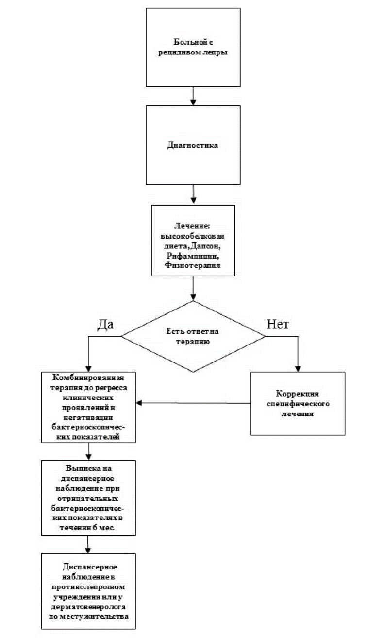
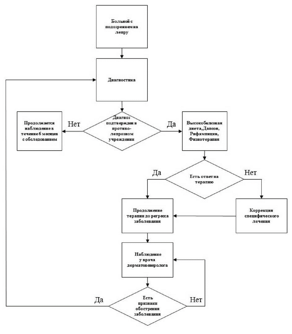 Приложение Б2. Алгоритмы действий врача (рецидив лепры)